European Society for Spatial Biology (ESSB)

European Society for Spatial Biology

Travel grants will be awarded for selected abstracts!

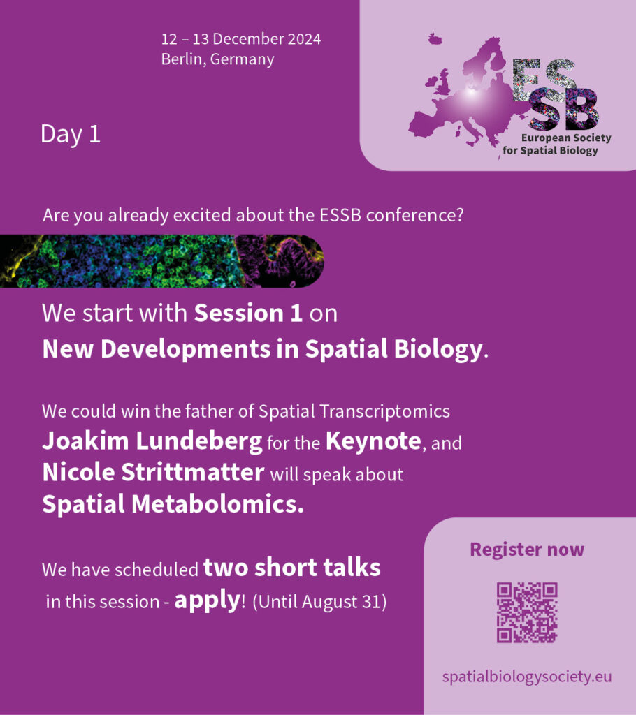
Great speaker line-up at the ESSB conference! Register and submit abstracts for short talks and posters until August 31st! 

Session 1 on December 12th:  @joalunatkthse and Nicole Strittmatter

#spatialbiology #spatialtranscriptomics #spatialmetabolomics #ESSB

New Developments in Spatial Biology

Chairs: Denis Schapiro, Christian Schürch

Thursday, 12.12.2024, 8:30 am

Joakim Lundeberg

Keynote Speaker: Joakim Lundeberg
SciLifeLab, KTH Royal Institute of Technology

Resolving the tissue ecosystem into its components

Tissue represents an ecosystem of different cells carrying out various tasks. Specific types of cells exist in every organ and serve specialized functions defined by the specific genes and proteins active in each cell type. Comprehensive maps of molecularly defined human cell types are underway through the Human Cell Atlas effort using primarily single-cell RNA sequencing. The technologies to assemble spatial maps that describe and explain the cellular basis of health and disease are still being discussed. We have developed and established the Spatial Transcriptomics technology, in which tissue imaging is merged with spatial RNA sequencing and resolved by computational means. Spatial Transcriptomics technology was the first method to provide unbiased whole transcriptome analysis with spatial information from tissue using barcoded array surfaces and has, since its initial publication, been used in multiple biological systems in health and disease. This presentation will cover the technology’s novel methodological and analytical aspects in the context of biological applications from cell atlas, neurology, and cancer.

Biosketch

Joakim Lundeberg, Ph.D., Professor in Molecular Biotechnology, was a co-founder of Science for Life Laboratory Sweden in 2010, a national and multi-university effort in large-scale life sciences providing access to infrastructures such as genomics, and proteomics, imaging, metabolomics, and drug development. Dr. Lundeberg has, during the most recent years, focused on spatial transcriptomics, which enables a detailed description of gene expression patterns in tissue sections. The methodology is now available worldwide through 10x Genomics Inc. The technology was also featured in Nature Methods as the Method of the Year 2020. Dr. Lundeberg has publications demonstrating technology development and examples of spatial analysis’s impact in biology. The current research focuses on expanding the spatial modalities and developing new software tools and applications in human cell atlas, neurology, and cancer.

Nicole Strittmatter

Invited talk by Nicole Strittmatter
Department of Bioscience, Technical University of Munich

Studying disease states in cancer and infection using spatial metabolomics

Mass Spectrometry Imaging (MSI) is a powerful technique to characterise complex biological samples such as tissues or plant material, enabling the mapping of hundreds of chemical constituents simultaneously. In the Strittmater lab, we are using MSI predominantly to map changes in the metabolome (spatial metabolomics) of biomedical specimen ranging from organoids to preclinical and clinical tissue specimen using Desorption Electrospray Ionisation (DESI) MSI, an ambient technique operating under atmospheric conditions and enabling analysis without prior sample preparation. This makes DESI-MSI particularly suited for deployment in multimodal imaging studies, such as in combination with imaging mass cytometry, which allows the targeted, multiplexed detection of proteins. In this talk, I will highlight some recent applications that cover some of our dominant research interests such as host-microbe interactions, tumour biology and anti-tumour drug disposition and metabolism.

Biosketch
Prof. Dr. Nicole Strittmatter is an expert in in situ mass spectrometry techniques, especially ambient mass spectrometry and imaging mass spectrometry. She has studied Chemistry at the Justus-Liebig University in Gießen, Germany, obtaining her MSc in 2011. She performed her PhD studies at the Department of Surgery and Cancer, Imperial College London, developing microbial identification tools using REIMS. Her postdoctoral years (2015-2021) were spent in industry, performing MS imaging studies at AstraZeneca, Cambridge, UK to study drug delivery and metabolism. Since 10.2021, she holds the assistant professorship of analytical chemistry (tenure track) at the Department of Biosciences, School of Natural Sciences, Technical University of Munich (TUM). Her current research focusses novel MS method development for spatial metabolomics and multimodal imaging in cancer, host-microbe interactions studies and beyond.


Short talk by Xavier Rovira Clavé

Department of Pediatrics,  Genetics, Stanford University, CA 94305, USA;

In Situ Tracking of Clonal Evolution and Phenotypic Heterogeneity in Tumors by Spatial Epitope Barcoding

Understanding the dynamics of clonal evolution and phenotypic shifts within the tumor microenvironment is crucial for developing effective therapeutic strategies. Previously, we introduced a combinatorial tagging system coupled with multiplexed ion beam imaging for in situ tracking of barcodes within tissues. This approach enabled us to dissect the spatial component of cell lineages and phenotypes in small cell lung cancer xenograft models, revealing emergent properties from mixed clones and the preferential expansion of clonal patches. We are now extending this approach by integrating it with genome editing technologies, allowing the investigation of the impact of genetic perturbations on clonal dynamics and phenotypic heterogeneity within the tumor microenvironment. We applied this integrated approach to study the effects of knocking out a set of key genes for small cell lung cancer development in a xenograft model. Our analysis revealed distinct patterns of clonal expansion and phenotypic shifts associated with specific gene knockouts and identified unique subclonal interactions within the tumor. This work highlights the complex interplay between genetic alterations, clonal dynamics, and phenotypic heterogeneity in the tumor microenvironment. Our findings demonstrate the potential of this approach to uncover novel insights into the mechanisms driving tumor progression and treatment resistance, paving the way for the development of more effective personalized therapies.

Short talk by Carsten Hopf

Center for Mass Spectrometry and Optical Spectroscopy (CeMOS), Mannheim University of Applied Sciences, Paul-Wittsack Str. 10, 68163, Mannheim, Germany.
Faculty of Medicine, Heidelberg University, Grabengasse 1, 69117 Heidelberg, Germany
Mannheim Center for Translational Neuroscience (MCTN), Medical Faculty Mannheim, Heidelberg University, 68167 Mannheim, Germany

Multimodal MALDI Lipid-Protein Imaging of Amyloid-β Plaques and Machine Learning reveal Alzheimer’s Disease Signatures

Alzheimer’s disease (AD), the leading cause of dementia, is characterized by deposition of amyloid-β peptides and various lipids in plaques. This study investigates the lipid and protein heterogeneity of amyloid-β (Aβ) plaques in the APP PS1 mouse model and in post-mortem brain samples from AD patients as well as in plaque-bearing but cognitively unimpaired individuals. We combined multimodal MALDI imaging of lipids and proteins at a single-plaque level on various mass spectrometry platforms with computational assessment of lipid and protein composition in large plaque populations. We identified distinct populations of amyloid plaques characterized by differential Aβ and lipid composition. These findings suggest that the heterogeneity in Aβ metabolism and lipid homeostasis is a key factor in the pathogenesis of AD and imply that total amyloid burden alone is an insufficient marker for the disease.

Brain sections of APP PS1 mice were analyzed by MALDI imaging on a neoflex benchtop MALDI-TOF mass spectrometer (Bruker). Lipid and HiPlex-MALDI-IHC information were obtained from the same tissue section. Multimodal MALDI lipid-protein imaging of fresh-frozen human brain sections was performed on a timsTOFflex mass spectrometer (Bruker). Computational evaluation of amyloid plaque-like objects was performed using the PlaquePicker R tool (Enzlein et al. 2020). Different machine learning approaches were utilized, in order to identify a mixed lipid-protein classifier characteristic for AD (Enzlein et al. 2024). 

Amyloid beta plaques were segmented using PlaquePicker and differentiated based on their lipid and peptide composition. Combination of MALDI lipid imaging and HiPLEX-IHC demonstrated extensive co-localization of gangliosides as well as with microglia and activated astrocytes with Aβ plaques. However, we identified only one class of plaques in the APP PS1 mouse model. In contrast, multimodal MALDI lipid-protein imaging of human brain samples followed by statistical single-plaque analysis and machine learning, revealed marked differences between plaques in AD tissue and in brains from plaque-bearing but cognitively unimpaired (AP-CU) individuals. Surprisingly, three different machine learning models suggested that Aβ1-38 and distinct gangliosides rather than Aβ1-42 which is believed to drive amyloid plaque formation, most strongly distinguishes between AD- and APCU amyloid plaques. The combination of these molecular markers in multimodal MS imaging could be used to better understand AD pathology.