European Society for Spatial Biology (ESSB)

European Society for Spatial Biology

Travel grants will be awarded for selected abstracts!

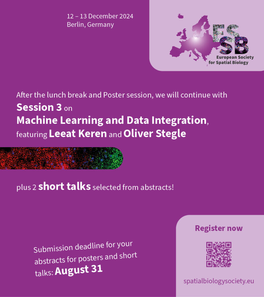
The 3rd session of the ESSB conference will be dedicated to spatial multi-omics, featuring Leeat Keren and Oliver Stegle as speakers. Again, there will also be presentations of selected abstracts!

Machine Learning and Data Integration in Spatial Biology

Chairs: Julio Saez-Rodriguez, Leeat Keren

Thursday, 12.12.2024, 2:00 pm

Invited talk by Leeat Keren
Molecular Cell Biology, Weizmann Institute of science, Rehovot, Israel

Escalating high-dimensional imaging for cancer research

Tumors are spatially organized ecosystems that are comprised of distinct cell types, each of which can assume a variety of phenotypes defined by coexpression of multiple proteins. To underscore this complexity it is essential to interrogate cellular expression patterns within their native context in the tissue. I will describe technological and computational advancements in multiplexed imaging, and demonstrate its application to study the tumor microenvironment.

Biosketch

Dr. Leeat Keren is an assistant professor in the department of Molecular Cell Biology, Weizmann Institute of science. She completed her Ph.D. in computational biology in the Weizmann Institute of Science in 2016 and her postdoc with Dr. Michael Angelo in Stanford University in 2020. She is an Azrieli Faculty Fellow, a recipient of the Dale F. Frey breakthrough scientist award from the Damon Runyon Cancer Research Foundation and a recipient of the James Heineman Research Award.


Oliver Stegle

Invited talk by Oliver Stegle

Computational Genomics & Systems Genetics, German Cancer Research Center (DKFZ), Heidelberg Germany

BioSketch

Prof. Dr. Oliver Stegle is the Head of the Computational Genomics and Systems Genetics Division at the German Cancer Research Center (DKFZ) and group leader at EMBL in Heidelberg, Germany. His laboratory is developing and applying statistical and machine learning methods for deciphering molecular variation across individuals, space and time. He coordinates the German Human-Genome-Phenome Archive, the health program of the European Laboratory for Learning and Intelligent Systems and is an ERC investigator.

Short talk by Lorenz Rumberger

Max-Delbruck-Center for Molecular Medicine in the Helmholtz Association, Berlin, Germany
Humboldt-Universität zu Berlin, Faculty of Mathematics and Natural Sciences, Berlin, Germany
Helmholtz Imaging

Automated classification of cellular expression in multiplexed imaging
data with Nimbus

Multiplexed imaging offers a powerful approach to characterize the spatial topography of tissues in both health and disease. To analyze such data, the specific combination of markers that are present in each cell must be enumerated in a process known as cell clustering. For clustering, the image data is transformed to tabular data, by integrating the marker expressions of the individual cell segments. This step introduces errors due to multiple confounders: signal can spill over from adjacent cells, uneven illumination and background staining can introduce false positives. To tackle this, we constructed the Pan-Multiplex (Pan-M) dataset containing 197 million distinct annotations of marker expression across 15 different cell types. We used Pan-M to create Nimbus, a deep learning model to predict marker positivity. Nimbus is a pre-trained model that can predict marker expression across distinct cell types, from different tissues, acquired using different microscope platforms, without requiring any retraining. We demonstrate that Nimbus predictions capture the underlying staining patterns of the full diversity of markers present in Pan-M. We then show how Nimbus predictions can be integrated with downstream clustering algorithms to robustly identify cell subtypes in image data. All underlying data, models, and code have been released as a community resource.

Short talk by André Rendeiro

CeMM Research Center for Molecular Medicine of the Austrian Academy of Sciences,
Lazarettgasse 14 AKH BT 25.3, 1090, Vienna, Austria
Ludwig Boltzmann Institute for Network Medicine at the University of Vienna, Vienna, Austria

Tissue-specific prediction of biological age from histopathological images

Aging, the leading risk factor for many diseases, manifests in diverse forms across human tissue architecture, providing an opportunity to quantify tissue-specific aging. To comprehensively understand the relationship between tissue structure, aging and pathology in humans, we present a comprehensive analysis of changes during aging using whole slide histopathological images from the Genotype-Tissue Expression Project (GTEx). Analyzing 25,712 images from 40 tissue types across 983 individuals, we apply deep learning to quantify age-related morphological changes.

We develop ‚tissue-clocks’—predictors of biological age from tissue images—achieving a mean prediction error of 4.9 years. These clocks correlate with telomere attrition, subclinical pathologies, and comorbidities. Assessing biological aging rates across organs reveals non-uniform aging patterns, with some organs showing early changes (20-40 years) and others displaying bimodal age-related patterns. We also identify associations between demographic, lifestyle, and medical history factors and tissue-specific acceleration or deceleration of biological age, highlighting modifiable risk factors.

This work bridges molecular and cellular changes to tissue-level contexts and demonstrates the value of histopathological imaging for understanding age-associated pathology in humans.